
Guide to Grading and Classification of Diabetic Retinopathy


What is Diabetic Retinopathy?
Diabetic retinopathy is a serious complication of diabetes that affects the retina: the light-sensitive tissue at the back of the eye. This condition develops when prolonged high blood sugar levels (hyperglycemia) damage the tiny blood vessels in the retina, leading to fluid accumulation in the retinal tissue.
The underlying mechanism involves the formation of advanced glycation end products (AGEs) that disrupt the blood-retinal barrier. This disruption compromises the integrity of endothelial cell junctions in retinal blood vessels, allowing fluid and other molecules to leak into the surrounding tissue.
Why Grading Matters
Proper grading of diabetic retinopathy is crucial for several reasons:
- Accurate diagnosis of disease severity
- Treatment planning based on risk stratification
- Disease monitoring to track progression over time
- Clinical decision-making regarding intervention timing
Healthcare providers typically assess diabetic retinopathy using fundoscopic examination, fundus photography, or optical coherence tomography (OCT) to visualize retinal changes.
The ETDRS Classification System
The standard grading system, established by the landmark Early Treatment Diabetic Retinopathy Study (ETDRS), categorizes diabetic retinopathy into five main stages:
1. No Diabetic Retinopathy
- No retinal abnormalities detected
2. Mild Non-Proliferative Diabetic Retinopathy (NPDR)
- Microaneurysms only
- These appear as tiny red dots on examination and represent small outpouchings of retinal capillaries - the earliest visible sign of diabetic retinopathy
3. Moderate Non-Proliferative Diabetic Retinopathy (NPDR)
- More extensive retinal changes, including:
- Increased number of microaneurysms
- Dot-blot hemorrhages
- Caused by blood leakage from damaged vessels
- Exudates
- Yellowish deposits resulting from fluid and lipoprotein leakage into the retina
- Cotton wool spots
- Indicates areas of retinal ischemia
In the opthamological world, there is no such thing as soft/hard exudates.
4. Severe Non-Proliferative Diabetic Retinopathy (NPDR)
- The "4-2-1 Rule" (presence of any one of the following):
- >20 intraretinal hemorrhages in each of the 4 retinal quadrants
- Venous beading in 2 or more quadrants
- Intraretinal microvascular abnormalities (IRMA) in 1 or more quadrants
- A/V shunts that run from the retina arterioles to the venules (bypassing the capillary bed)
- Significant capillary occlusion (drop-out)
5. Proliferative Diabetic Retinopathy (PDR)
- Neovascularization (formation of new, abnormal blood vessels on the retina or optic disc)
- Vitreous or preretinal hemorrhage
- Fibrous tissue proliferation
- Tractional retinal detachment due to scar tissue formation
- High risk (Any one of the following)
- Neovascularization of the disc (NVD) > 1/4 to 1/3 disc area
- Any NVD associated with vitreous or preretinal hemorrhage
- Any neovascularization elsewhere (NVE) associated with vitreous or preretinal hemorrhage
Diabetic Macular Edema
Diabetic Macular Edema (DME) requires separate classification because it can occur at any stage of diabetic retinopathy and specifically affects the macula: the central part of the retina responsible for sharp, detailed vision.
DME Classification:
- Absent:
- No macular involvement
- Present: Further categorized as:
- Focal thickening of the macula
- Diffuse thickening of the macula
- Clinically significant macular edema (CSME) when meeting specific proximity and severity criteria
| Stage | Key Features |
|---|---|
| No DR | Normal retina |
| Mild NPDR | Microaneurysms only |
| Moderate NPDR | More microaneurysms, hemorrhages, hard exudates |
| Severe NPDR | 4-2-1 rule criteria met, no neovascularization |
| Proliferative DR (PDR) | Neovascularization present, risk of hemorrhage and retinal detachment |
| Diabetic Macular Edema (DME) | Can occur at any stage; specifically affects central vision |
Treatment Recommendations
Published by the AAO, the following table describes the best treatment recommendations and follow-up duration for the initial management of patients with diabetes.
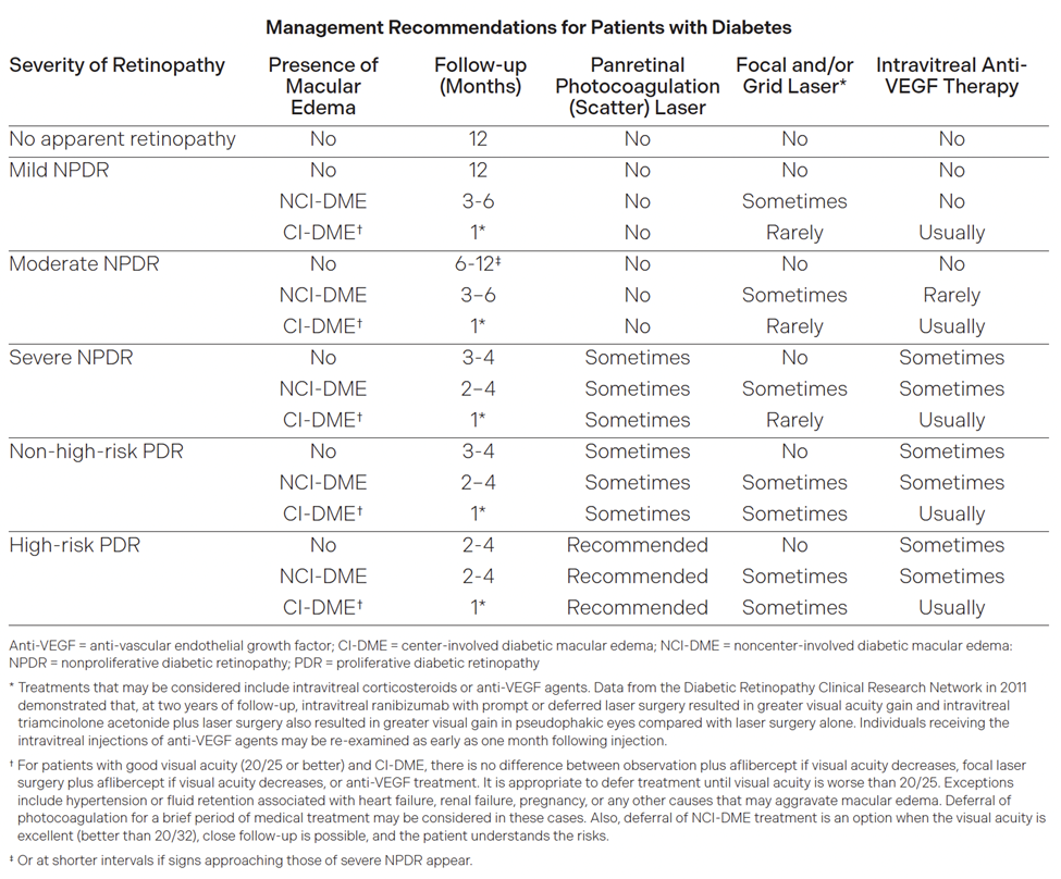
All center invloved DME (CI-DME) have a 1mo follow up regardless of their grade.